Registro completo de metadatos
| Campo DC | Valor | Lengua/Idioma |
|---|---|---|
| dc.creator | Vicent, S. (Silvestre) | |
| dc.creator | Lopez-Picazo, J.M. (José M.) | |
| dc.creator | Toledo, G. (Gemma) | |
| dc.creator | Lozano, M.D. (María Dolores) | |
| dc.creator | Torre, W. (Wenceslao) | |
| dc.creator | Garcia-Corchon, C. (C.) | |
| dc.creator | Quero, C. (C.) | |
| dc.creator | Soria, J.C. (Jean-Charles) | |
| dc.creator | Martin-Algarra, S. (Salvador) | |
| dc.creator | Manzano, R.G. (Ramón G.) | |
| dc.creator | Montuenga-Badia, L.M. (Luis M.) | |
| dc.date.accessioned | 2011-08-24T07:11:53Z | - |
| dc.date.available | 2011-08-24T07:11:53Z | - |
| dc.date.issued | 2004 | - |
| dc.identifier.citation | Vicent S, Lopez-Picazo JM, Toledo G, Lozano MD, Torre W, Garcia-Corchon C, et al. ERK1/2 is activated in non-small-cell lung cancer and associated with advanced tumours. Br J Cancer 2004 Mar 8;90(5):1047-1052. | es_ES |
| dc.identifier.issn | 1532-1827 | - |
| dc.identifier.uri | https://hdl.handle.net/10171/18817 | - |
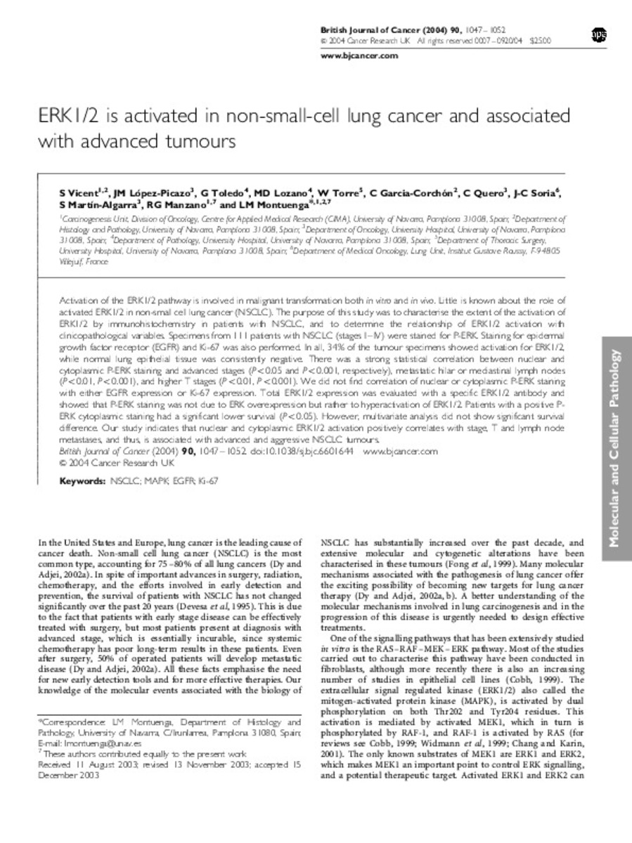
| dc.description.abstract | Activation of the ERK1/2 pathway is involved in malignant transformation both in vitro and in vivo. Little is known about the role of activated ERK1/2 in non-small cell lung cancer (NSCLC). The purpose of this study was to characterise the extent of the activation of ERK1/2 by immunohistochemistry in patients with NSCLC, and to determine the relationship of ERK1/2 activation with clinicopathological variables. Specimens from 111 patients with NSCLC (stages I-IV) were stained for P-ERK. Staining for epidermal growth factor receptor (EGFR) and Ki-67 was also performed. In all, 34% of the tumour specimens showed activation for ERK1/2, while normal lung epithelial tissue was consistently negative. There was a strong statistical correlation between nuclear and cytoplasmic P-ERK staining and advanced stages (P<0.05 and P<0.001, respectively), metastatic hilar or mediastinal lymph nodes (P<0.01, P<0.001), and higher T stages (P<0.01, P<0.001). We did not find correlation of nuclear or cytoplasmic P-ERK staining with either EGFR expression or Ki-67 expression. Total ERK1/2 expression was evaluated with a specific ERK1/2 antibody and showed that P-ERK staining was not due to ERK overexpression but rather to hyperactivation of ERK1/2. Patients with a positive P-ERK cytoplasmic staining had a significant lower survival (P<0.05). However, multivariate analysis did not show significant survival difference. Our study indicates that nuclear and cytoplasmic ERK1/2 activation positively correlates with stage, T and lymph node metastases, and thus, is associated with advanced and aggressive NSCLC tumours. | es_ES |
| dc.language.iso | eng | es_ES |
| dc.publisher | Nature Publishing Group | es_ES |
| dc.rights | info:eu-repo/semantics/openAccess | es_ES |
| dc.subject | NSCLC | es_ES |
| dc.subject | MAPK | es_ES |
| dc.subject | EGFR | es_ES |
| dc.subject | Ki-67 | es_ES |
| dc.title | ERK1/2 is activated in non-small-cell lung cancer and associated with advanced tumours | es_ES |
| dc.type | info:eu-repo/semantics/article | es_ES |
| dc.relation.publisherversion | http://www.nature.com/bjc/journal/v90/n5/full/6601644a.html | es_ES |
Ficheros en este ítem:
Estadísticas e impacto
Los ítems de Dadun están protegidos por copyright, con todos los derechos reservados, a menos que se indique lo contrario.


