Full metadata record
| DC Field | Value | Language |
|---|---|---|
| dc.creator | Becerra, S.P. (S.P.) | - |
| dc.creator | Fariss, R.N. (R. N.) | - |
| dc.creator | Wu, Y.Q. (Yan Q.) | - |
| dc.creator | Montuenga-Badia, L.M. (Luis M.) | - |
| dc.creator | Wong, P. (Paul) | - |
| dc.creator | Pfeffer, B.A. (Bruce A.) | - |
| dc.date.accessioned | 2011-12-12T08:52:21Z | - |
| dc.date.available | 2011-12-12T08:52:21Z | - |
| dc.date.issued | 2004 | - |
| dc.identifier.citation | Becerra SP, Fariss RN, Wu YQ, Montuenga LM, Wong P, Pfeffer BA. Pigment epithelium-derived factor in the monkey retinal pigment epithelium and interphotoreceptor matrix: apical secretion and distribution. Exp Eye Res 2004 Feb;78(2):223-234. | es_ES |
| dc.identifier.issn | 1096-0007 | - |
| dc.identifier.uri | https://hdl.handle.net/10171/20178 | - |
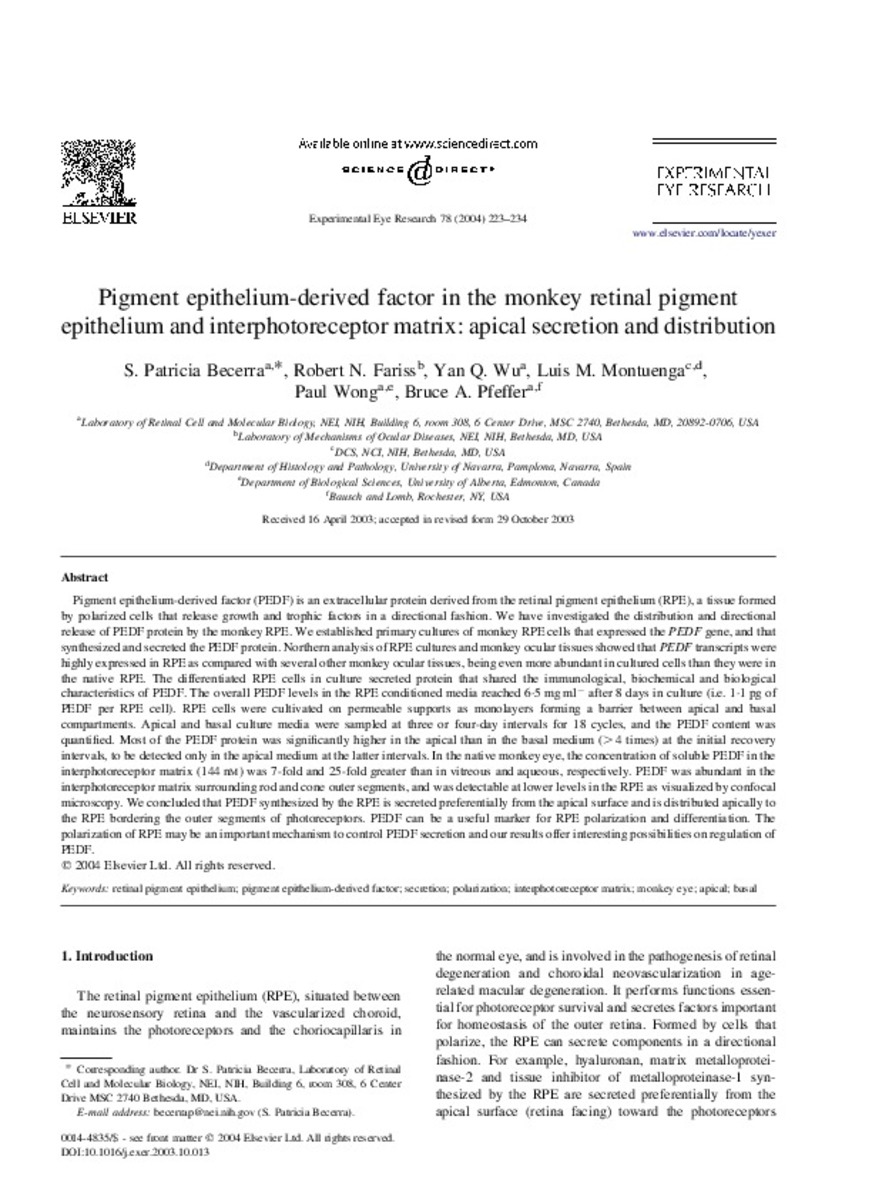
| dc.description.abstract | Pigment epithelium-derived factor (PEDF) is an extracellular protein derived from the retinal pigment epithelium (RPE), a tissue formed by polarized cells that release growth and trophic factors in a directional fashion. We have investigated the distribution and directional release of PEDF protein by the monkey RPE. We established primary cultures of monkey RPE cells that expressed the PEDF gene, and that synthesized and secreted the PEDF protein. Northern analysis of RPE cultures and monkey ocular tissues showed that PEDF transcripts were highly expressed in RPE as compared with several other monkey ocular tissues, being even more abundant in cultured cells than they were in the native RPE. The differentiated RPE cells in culture secreted protein that shared the immunological, biochemical and biological characteristics of PEDF. The overall PEDF levels in the RPE conditioned media reached 6.5 mg ml- after 8 days in culture (i.e. 1.1 pg of PEDF per RPE cell). RPE cells were cultivated on permeable supports as monolayers forming a barrier between apical and basal compartments. Apical and basal culture media were sampled at three or four-day intervals for 18 cycles, and the PEDF content was quantified. Most of the PEDF protein was significantly higher in the apical than in the basal medium (>4 times) at the initial recovery intervals, to be detected only in the apical medium at the latter intervals. In the native monkey eye, the concentration of soluble PEDF in the interphotoreceptor matrix (144 nM) was 7-fold and 25-fold greater than in vitreous and aqueous, respectively. PEDF was abundant in the interphotoreceptor matrix surrounding rod and cone outer segments, and was detectable at lower levels in the RPE as visualized by confocal microscopy. We concluded that PEDF synthesized by the RPE is secreted preferentially from the apical surface and is distributed apically to the RPE bordering the outer segments of photoreceptors. PEDF can be a useful marker for RPE polarization and differentiation. The polarization of RPE may be an important mechanism to control PEDF secretion and our results offer interesting possibilities on regulation of PEDF. | es_ES |
| dc.language.iso | eng | es_ES |
| dc.publisher | Elsevier | es_ES |
| dc.rights | info:eu-repo/semantics/openAccess | es_ES |
| dc.subject | Retinal pigment epithelium | es_ES |
| dc.subject | Pigment epithelium-derived factor | es_ES |
| dc.subject | Secretion | es_ES |
| dc.subject | Polarization | es_ES |
| dc.subject | Interphotoreceptor matrix | es_ES |
| dc.subject | Monkey eye | es_ES |
| dc.subject | Apical | es_ES |
| dc.subject | Basal | es_ES |
| dc.title | Pigment epithelium-derived factor in the monkey retinal pigment epithelium and interphotoreceptor matrix: apical secretion and distribution | es_ES |
| dc.type | info:eu-repo/semantics/article | es_ES |
| dc.relation.publisherversion | http://www.sciencedirect.com/science/article/pii/S0014483503003610 | es_ES |
| dc.type.driver | info:eu-repo/semantics/article | es_ES |
Files in This Item:
Statistics and impact
Items in Dadun are protected by copyright, with all rights reserved, unless otherwise indicated.


