Full metadata record
| DC Field | Value | Language |
|---|---|---|
| dc.creator | Alava, E. (Enrique) de | - |
| dc.creator | Panizo, A. (Ángel) | - |
| dc.creator | Antonescu, C.R. (Cristina R.) | - |
| dc.creator | Huvos, A.G. (Andrew G.) | - |
| dc.creator | Pardo-Mindan, F.J. (Francisco Javier) | - |
| dc.creator | Barr, F.G. (Frederic G.) | - |
| dc.creator | Ladanyi, M. (Marc) | - |
| dc.date.accessioned | 2012-10-16T08:51:02Z | - |
| dc.date.available | 2012-10-16T08:51:02Z | - |
| dc.date.issued | 2000 | - |
| dc.identifier.citation | de Alava E, Panizo A, Antonescu CR, Huvos AG, Pardo-Mindan FJ, Barr FG, et al. Association of EWS-FLI1 type 1 fusion with lower proliferative rate in Ewing's sarcoma. Am J Pathol 2000 Mar;156(3):849-855. | es_ES |
| dc.identifier.issn | 0002-9440 | - |
| dc.identifier.uri | https://hdl.handle.net/10171/23392 | - |
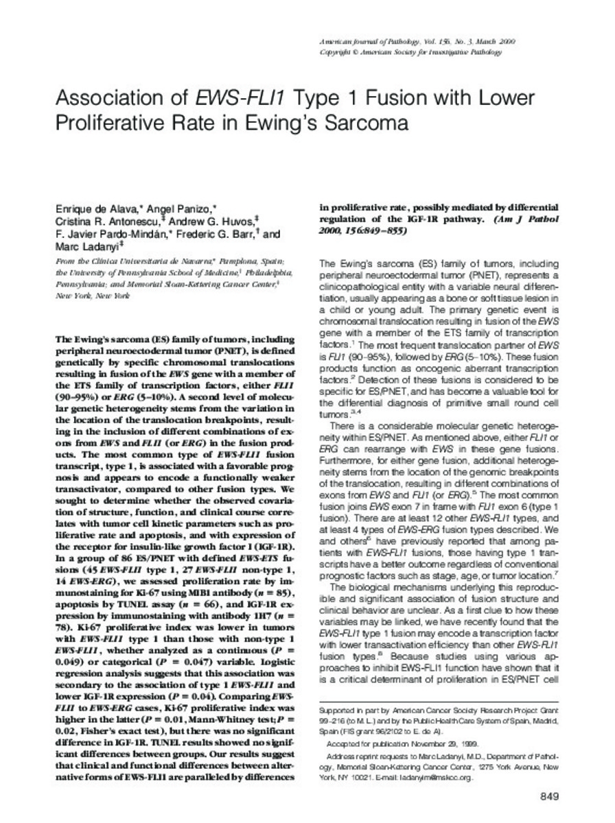
| dc.description.abstract | The Ewing's sarcoma (ES) family of tumors, including peripheral neuroectodermal tumor (PNET), is defined genetically by specific chromosomal translocations resulting in fusion of the EWS gene with a member of the ETS family of transcription factors, either FLI1 (90-95%) or ERG (5-10%). A second level of molecular genetic heterogeneity stems from the variation in the location of the translocation breakpoints, resulting in the inclusion of different combinations of exons from EWS and FLI1 (or ERG) in the fusion products. The most common type of EWS-FLI1 fusion transcript, type 1, is associated with a favorable prognosis and appears to encode a functionally weaker transactivator, compared to other fusion types. We sought to determine whether the observed covariation of structure, function, and clinical course correlates with tumor cell kinetic parameters such as proliferative rate and apoptosis, and with expression of the receptor for insulin-like growth factor I (IGF-1R). In a group of 86 ES/PNET with defined EWS-ETS fusions (45 EWS-FLI1 type 1, 27 EWS-FLI1 non-type 1, 14 EWS-ERG), we assessed proliferation rate by immunostaining for Ki-67 using MIB1 antibody (n = 85), apoptosis by TUNEL assay (n = 66), and IGF-1R expression by immunostaining with antibody 1H7 (n = 78). Ki-67 proliferative index was lower in tumors with EWS-FLI1 type 1 than those with non-type 1 EWS-FLI1, whether analyzed as a continuous (P = 0.049) or categorical (P = 0.047) variable. Logistic regression analysis suggests that this association was secondary to the association of type 1 EWS-FLI1 and lower IGF-1R expression (P = 0.04). Comparing EWS-FLI1 to EWS-ERG cases, Ki-67 proliferative index was higher in the latter (P = 0.01, Mann-Whitney test; P = 0.02, Fisher's exact test), but there was no significant difference in IGF-1R. TUNEL results showed no significant differences between groups. Our results suggest that clinical and functional differences between alternative forms of EWS-FLI1 are paralleled by differences in proliferative rate, possibly mediated by differential regulation of the IGF-1R pathway. | es_ES |
| dc.language.iso | eng | es_ES |
| dc.publisher | Elsevier | es_ES |
| dc.rights | info:eu-repo/semantics/openAccess | es_ES |
| dc.subject | DNA-Binding Proteins | es_ES |
| dc.subject | Bone Neoplasms/metabolism/pathology | es_ES |
| dc.subject | Immunoenzyme Techniques | es_ES |
| dc.title | Association of EWS-FLI1 Type 1 Fusion with Lower Proliferative Rate in Ewing’s Sarcoma | es_ES |
| dc.type | info:eu-repo/semantics/article | es_ES |
| dc.relation.publisherversion | http://www.ncbi.nlm.nih.gov/pmc/articles/PMC1876855/pdf/2107.pdf | es_ES |
| dc.type.driver | info:eu-repo/semantics/article | es_ES |
Files in This Item:
Statistics and impact
Items in Dadun are protected by copyright, with all rights reserved, unless otherwise indicated.


