Jejunal microvilli atrophy and reduced nutrient transport in rats with advanced liver cirrhosis: improvement by Insulin-like Growth Factor I
Palabras clave : 
Galactose/pharmacokinetics
Insulin-Like Growth Factor I/metabolism
Liver Cirrhosis/chemically induced/metabolism/pathology
Fecha de publicación : 
2004
Editorial : 
BioMed Central
ISSN : 
1471-230X
Cita: 
Castilla-Cortazar I, Pascual M, Urdaneta E, Pardo J, Puche JE, Vivas B, et al. Jejunal microvilli atrophy and reduced nutrient transport in rats with advanced liver cirrhosis: improvement by Insulin-like Growth Factor I. BMC Gastroenterol 2004 Jun 14;4:12.
Resumen
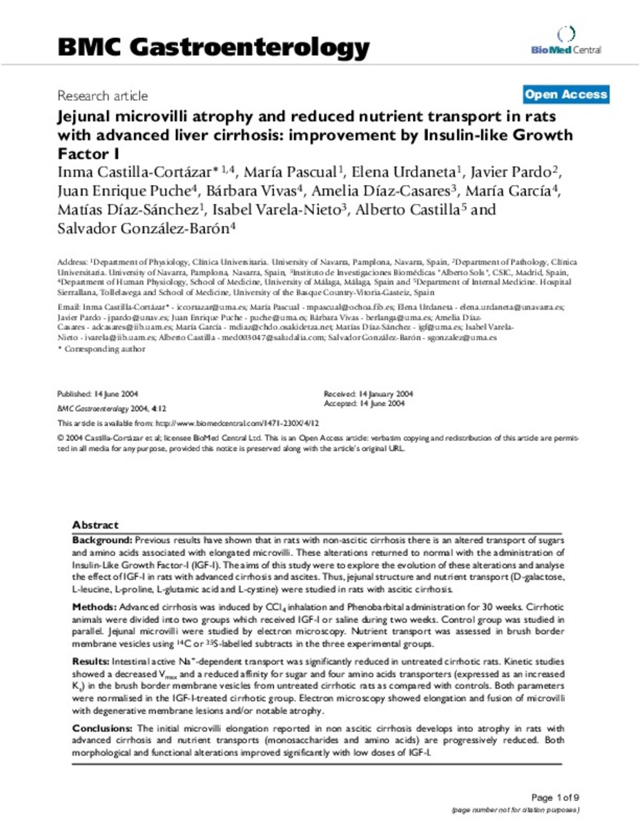
Previous results have shown that in rats with non-ascitic cirrhosis there is an altered transport of sugars and amino acids associated with elongated microvilli. These alterations returned to normal with the administration of Insulin-Like Growth Factor-I (IGF-I). The aims of this study were to explore the evolution of these alterations and analyse the effect of IGF-I in rats with advanced cirrhosis and ascites. Thus, jejunal structure and nutrient transport (D-galactose, L-leucine, L-proline, L-glutamic acid and L-cystine) were studied in rats with ascitic cirrhosis. METHODS: Advanced cirrhosis was induced by CCl4 inhalation and Phenobarbital administration for 30 weeks. Cirrhotic animals were divided into two groups which received IGF-I or saline during two weeks. Control group was studied in parallel. Jejunal microvilli were studied by electron microscopy. Nutrient transport was assessed in brush border membrane vesicles using 14C or 35S-labelled subtracts in the three experimental groups. RESULTS: Intestinal active Na+-dependent transport was significantly reduced in untreated cirrhotic rats. Kinetic studies showed a decreased Vmax and a reduced affinity for sugar and four amino acids transporters (expressed as an increased Kt) in the brush border membrane vesicles from untreated cirrhotic rats as compared with controls. Both parameters were normalised in the IGF-I-treated cirrhotic group. Electron microscopy showed elongation and fusion of microvilli with degenerative membrane lesions and/or notable atrophy. CONCLUSIONS: The initial microvilli elongation reported in non ascitic cirrhosis develops into atrophy in rats with advanced cirrhosis and nutrient transports (monosaccharides and amino acids) are progressively reduced. Both morphological and functional alterations improved significantly with low doses of IGF-I.

Ficheros en este ítem:
Vista previa
Fichero
BMC Gastroenterology 2004.12.pdf
Descripción
Tamaño
590.18 kB
Formato
Adobe PDF


Estadísticas e impacto
0 citas en
0 citas en

Los ítems de Dadun están protegidos por copyright, con todos los derechos reservados, a menos que se indique lo contrario.