PEGylated-PLGA microparticles containing VEGF for long term drug delivery
Palabras clave : 
PEG
PLGA
Macrophage uptake
VEGF
Protein delivery
Fecha de publicación : 
2013
Editorial : 
Elsevier
ISSN : 
0378-5173
Cita: 
Simon-Yarza T, Formiga FR, Tamayo E, Pelacho B, Prosper F, Blanco-Prieto MJ. PEGylated-PLGA microparticles containing VEGF for long term drug delivery. Int J Pharm 2013 JAN 2;440(1):13-18
Resumen
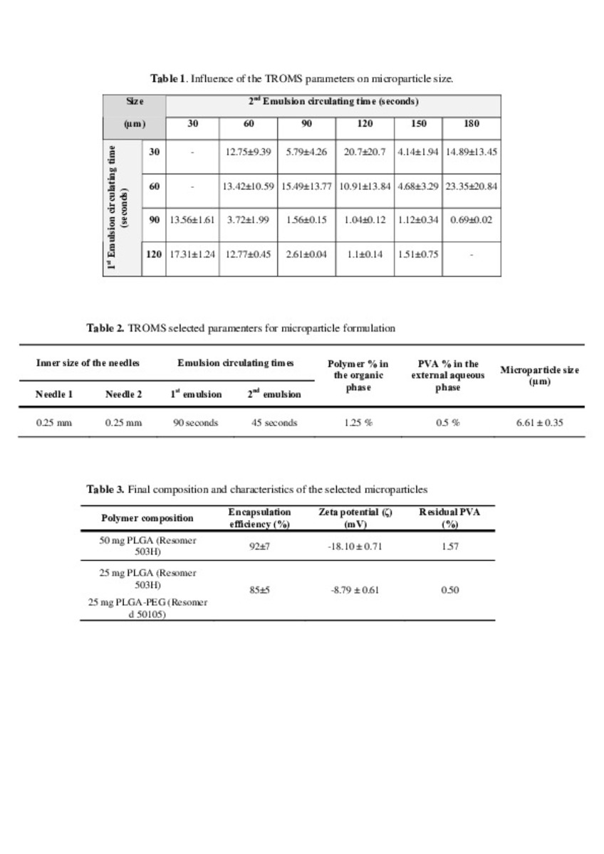
The potential of poly(lactic-co-glycolic) acid (PLGA) microparticles as carriers for vascular endothelial growth factor (VEGF) has been demonstrated in a previous study by our group, where we found improved angiogenesis and heart remodeling in a rat myocardial infarction model (Formiga et al., 2010). However, the observed accumulation of macrophages around the injection site suggested that the efficacy of treatment could be reduced due to particle phagocytosis. The aim of the present study was to decrease particle phagocytosis and consequently improve protein delivery using stealth technology. PEGylated microparticles were prepared by the double emulsion solvent evaporation method using TROMS (Total Recirculation One Machine System). Before the uptake studies in monocyte-macrophage cells lines (J774 and Raw 264.7), the characterization of the microparticles developed was carried out in terms of particle size, encapsulation efficiency, protein stability, residual poly(vinyl alcohol) (PVA) and in vitro release. Microparticles of suitable size for intramyocardial injection (5 mu m) were obtained by TROMS by varying the composition of the formulation and TROMS conditions with high encapsulation efficiency (70-90%) and minimal residual PVA content (0.5%). Importantly, the bioactivity of the protein was fully preserved. Moreover, PEGylated microparticles released in phosphate buffer 50% of the entrapped protein within 4 h, reaching a plateau within the first day of the in vitro study. Finally, the use of PLGA microparticles coated with PEG resulted in significantly decreased uptake of the carriers by macrophages, compared with non PEGylated microparticles, as shown by flow cytometry and fluorescence microscopy. On the basis of these results, we concluded that PEGylated microparticles loaded with VEGF could be used for delivering growth factors in the myocardium.

Ficheros en este ítem:
Vista previa
Fichero
Simon at al-Figure 3.tif
Descripción
Tamaño
337 kB
Formato
TIFF
Vista previa
Fichero
Simon et al-Figure 1.tif
Descripción
Tamaño
625.1 kB
Formato
TIFF
Vista previa
Fichero
Simon et al-Figure 2.tif
Descripción
Tamaño
108.07 kB
Formato
TIFF
Vista previa
Fichero
Simon et al-Figure 4.tif
Descripción
Tamaño
79.21 kB
Formato
TIFF
Vista previa
Fichero
Simon et al-Tables.pdf
Descripción
Tamaño
58.46 kB
Formato
Adobe PDF
Vista previa
Fichero
Simon et al-Manuscript.pdf
Descripción
Tamaño
138.88 kB
Formato
Adobe PDF


Estadísticas e impacto
0 citas en
0 citas en

Los ítems de Dadun están protegidos por copyright, con todos los derechos reservados, a menos que se indique lo contrario.