Characterization of Brucella abortus and Brucella melitensis native haptens as outer membrane O-type polysaccharides independent from the smooth lipopolysaccharide
Palabras clave : 
Radial immunodiffusion test
Escherichia-coli
Immunochemical characterization
Polyacrylamide gels
Bovine brucellosis
Silver stain
Antigen
Protein
Chain
Diagnosis
Fecha de publicación : 
1996
Editorial : 
American Society for Microbiology
ISSN : 
0021-9193
Cita: 
Aragon V, Diaz R, Moreno E, Moriyon I. Characterization of Brucella abortus and Brucella melitensis native haptens as outer membrane O-type polysaccharides independent from the smooth lipopolysaccharide. J Bacteriol 1996 Feb;178(4):1070-1079.
Resumen
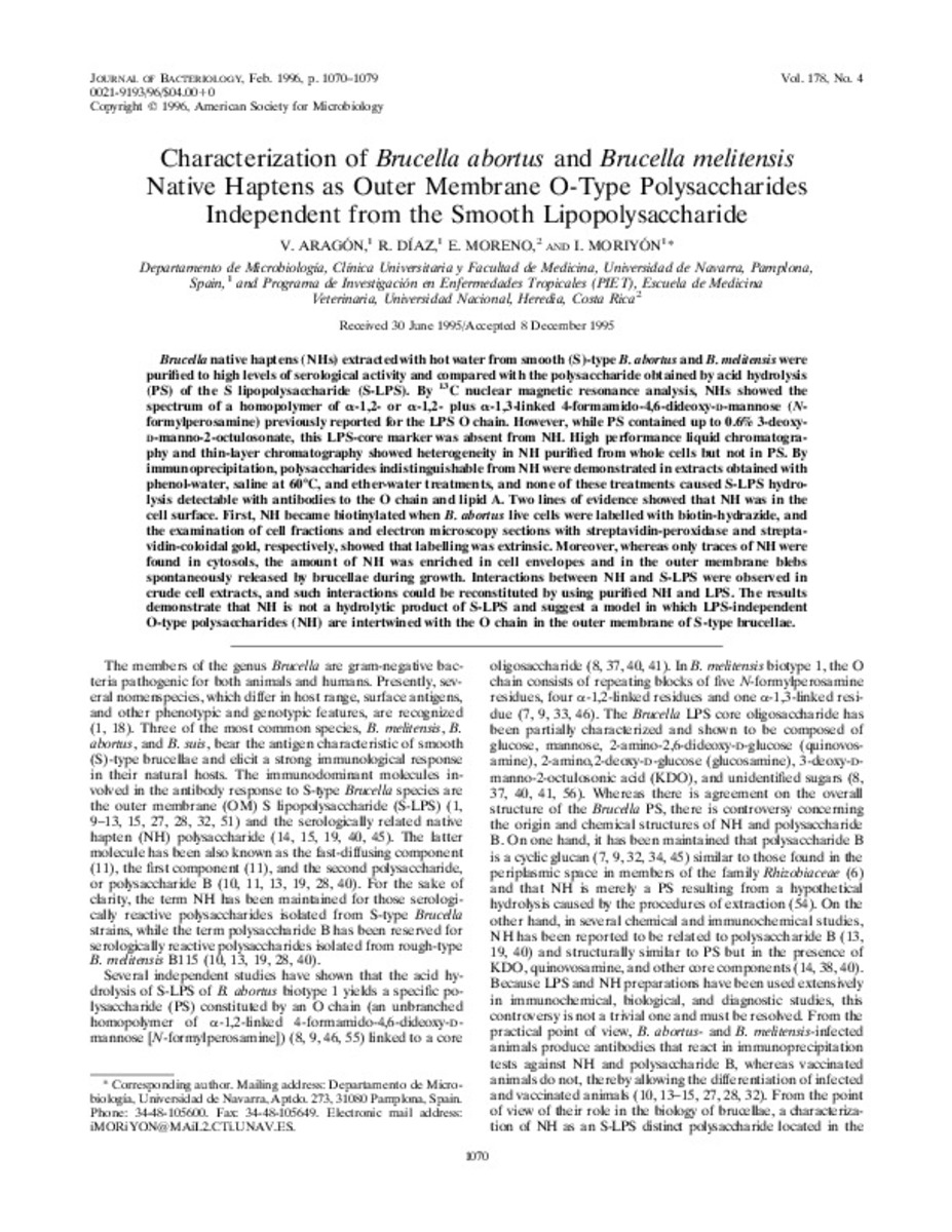
Brucella native haptens (NHs) extracted with hot water from smooth (S)-type B. abortus and B. melitensis were purified to high levels of serological activity and compared with the polysaccharide obtained by acid hydrolysis (PS) of the S lipopolysaccharide (S-LPS). By C-13 nuclear magnetic resonance analysis, NHs showed the spectrum of a homopolymer of alpha-1,2- or alpha-1-1,2- plus alpha-1,3-linked 4-formamido-4,6-dideoxy-D-mannose (N-formylperosamine) previously reported for the LPS O chain. However, while PS contained up to 0.6% 3-deoxy-D-manno-2-octulosonate, this LPS-core marker was absent from NH. High performance liquid chromatography and thin-layer chromatography showed heterogeneity in NH purified from whole cells but not in PS. By immunoprecipitation, polysaccharides indistinguishable from NH were demonstrated in extracts obtained with phenol-water, saline at 60 degrees C, and ether-water treatments, and none of these treatments caused S-LPS hydrolysis detectable with antibodies to the O chain and lipid A. Two lines of evidence showed that NH was in the cell surface. First, NH became biotinylated when B. abortus live cells were labelled with biotin-hydrazide, and the examination of cell fractions and electron microscopy sections with streptavidin-peroxidase and streptavidin-coloidal gold, respectively, showed that labelling was extrinsic. Moreover, whereas only traces of NH were found in cytosols, the amount of NH was enriched in cell envelopes and in the outer membrane blebs spontaneously released by brucellae during growth. Interactions between NH and S-LPS were observed in crude cell extracts, and such interactions could be reconstituted by using purified NH and LPS. The results demonstrate that NH is not a hydrolytic product of S-LPS and suggest a model in which LPS-independent O-type polysaccharides (NH) are intertwined with the O chain in the outer membrane of S-type brucellae.

Ficheros en este ítem:
Vista previa
Fichero
Characterization.pdf
Descripción
Tamaño
585.51 kB
Formato
Adobe PDF


Estadísticas e impacto
0 citas en
0 citas en

Los ítems de Dadun están protegidos por copyright, con todos los derechos reservados, a menos que se indique lo contrario.