Full metadata record
| DC Field | Value | Language |
|---|---|---|
| dc.creator | Ho, C.Y. (Carolyn Y.) | es_ES |
| dc.creator | Abbasi, S.A. (S.A.) | es_ES |
| dc.creator | Neilan, T.G. (T.G.) | es_ES |
| dc.creator | Shah, R.V.(R.V.) | es_ES |
| dc.creator | Chen, Y.(Y.) | es_ES |
| dc.creator | Heydari, B. (B.) | es_ES |
| dc.creator | Cirino, A.L. (Alison L.) | es_ES |
| dc.creator | Lakdawala, N.K. (Neal K.) | es_ES |
| dc.creator | Orav, E.J. (E. J.) | es_ES |
| dc.creator | González-Miqueo, A. (Aránzazu) | es_ES |
| dc.creator | Lopez-Salazar, M.B. (María Begoña) | es_ES |
| dc.creator | Diez-Martinez, J. (Javier) | es_ES |
| dc.creator | Jerosch-Herold, M. (M.) | es_ES |
| dc.creator | Kwong, R.Y. (Raymond Y.) | es_ES |
| dc.date.accessioned | 2014-05-30T11:42:33Z | - |
| dc.date.available | 2014-05-30T11:42:33Z | - |
| dc.date.issued | 2013 | es_ES |
| dc.identifier.citation | Ho, C. Y.; Abbasi, S. A.; Neilan, T. G.; Shah, R. V.; Chen, Y.; Heydari, B.; Cirino, A. L.; Lakdawala, N. K.; Orav, E. J.; González-Miqueo, A. ; López-Salazar, M. ; Díez-Martínez, D. ; Jerosch-Herold, M.; Kwong, R. Y. ""T1 measurements identify extracellular volume expansion in hypertrophic cardiomyopathy sarcomere mutation carriers with and without left ventricular hypertrophy ".Circulation: Cardiovasc Imaging. 2013 May 1; 6(3): 415–422 | en_EN |
| dc.identifier.issn | 1941-9651 | en_EN |
| dc.identifier.uri | https://hdl.handle.net/10171/35965 | - |
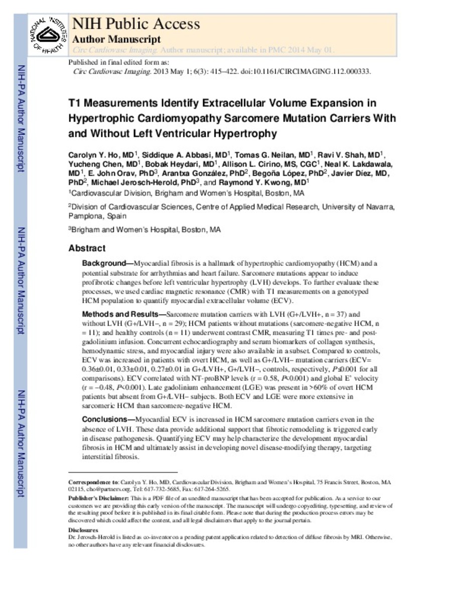
| dc.description.abstract | Background—Myocardial fibrosis is a hallmark of hypertrophic cardiomyopathy (HCM) and a potential substrate for arrhythmias and heart failure. Sarcomere mutations seem to induce profibrotic changes before left ventricular hypertrophy (LVH) develops. To further evaluate these processes, we used cardiac magnetic resonance with T1 measurements on a genotyped HCM population to quantify myocardial extracellular volume (ECV). Methods and Results—Sarcomere mutation carriers with LVH (G+/LVH+, n=37) and without LVH (G+/LVH−, n=29), patients with HCM without mutations (sarcomere-negative HCM, n=11), and healthy controls (n=11) underwent contrast cardiac magnetic resonance, measuring T1 times pre- and postgadolinium infusion. Concurrent echocardiography and serum biomarkers of collagen synthesis, hemodynamic stress, and myocardial injury were also available in a subset. Compared with controls, ECV was increased in patients with overt HCM, as well as G+/LVH− mutation carriers (ECV=0.36±0.01, 0.33±0.01, 0.27±0.01 in G+/LVH+, G+/LVH−, controls, respectively; P≤0.001 for all comparisons). ECV correlated with N-terminal probrain natriuretic peptide levels (r=0.58; P<0.001) and global E’ velocity (r=−0.48; P<0.001). Late gadolinium enhancement was present in >60% of overt patients with HCM but absent from G+/LVH− subjects. Both ECV and late gadolinium enhancement were more extensive in sarcomeric HCM than sarcomere-negative HCM. Conclusions—Myocardial ECV is increased in HCM sarcomere mutation carriers even in the absence of LVH. These data provide additional support that fibrotic remodeling is triggered early in disease pathogenesis. Quantifying ECV may help characterize the development of myocardial fibrosis in HCM and ultimately assist in developing novel disease-modifying therapy, targeting interstitial fibrosis. | en_EN |
| dc.language.iso | eng | en_EN |
| dc.rights | info:eu-repo/semantics/openAccess | es_ES |
| dc.subject | Fibrosis | en_EN |
| dc.subject | Hypertrophic cardiomyopath | es_ES |
| dc.subject | Genetic | en_EN |
| dc.subject | Magnetic resonance imaging | en_EN |
| dc.title | T1 measurements identify extracellular volume expansion in hypertrophic cardiomyopathy sarcomere mutation carriers with and without left ventricular hypertrophy | es_ES |
| dc.type | info:eu-repo/semantics/article | es_ES |
| dc.identifier.doi | http://dx.doi.org/10.1161/CIRCIMAGING.112.000333 | es_ES |
Files in This Item:
Statistics and impact
Items in Dadun are protected by copyright, with all rights reserved, unless otherwise indicated.


