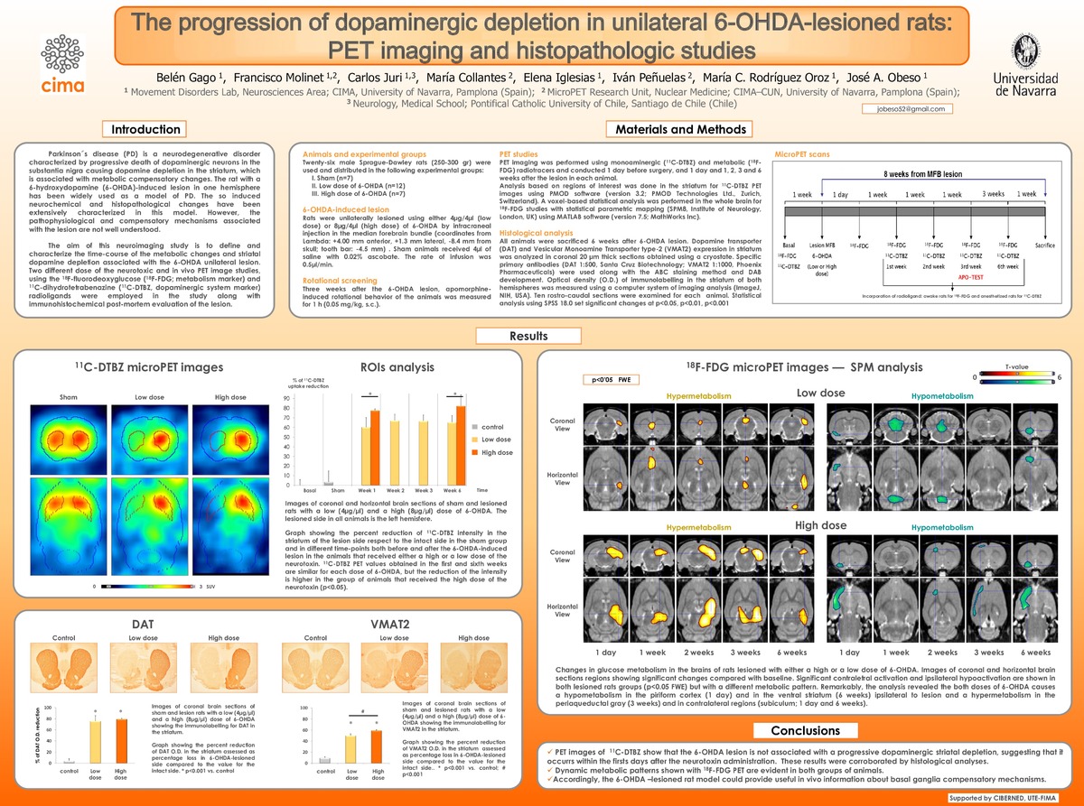
The progression of dopaminergic depletion in unilateral 6-OHDA-lesioned rats: PET imaging and histopathologic studies. | 8th FENS Forum of Neuroscience (14-18 July 2012. Barcelona, Spain)
Keywords: 
Rat Brain
18F-FDG
11C-DTBZ
Positron Emission Tomography
6-hydroxidopamine
Parkinson’s Disease
Issue Date: 
15-Jul-2012
Project: 
CIBERNED, UTE-FIMA
Abstract
Parkinson´s disease(PD) is characterize by a progressive death of dopaminergic neurons in the substantia nigra causing a dopamine depletion in the striatum, which is associated with metabolic compensatory changes. The rat with 6-hydroxydopamine (6-OHDA)-induced lesion in one hemisphere has been widely used as a model of PD. However, the series of pathophysiological and compensatory mechanisms associated with the lesion are not well understood. We performed a neuroimaging study aiming to define the functional changes associated with dopamine striatal depletion. Sprague-Dawley rats were unilaterally lesion using 4µg/4µl and 8µg/4µl of 6-OHDA by intracraneal injection in the median forebrain bundle. PET imaging was performed using a monoaminergic (11C-Dihydrotetrabenazine; 11C-DTBZ) and a metabolic (18F-fluorodeoxyglucose; 18F-FDG) radiotracer and conducted 1 day and 1, 2 , 3 and 6 weeks after the lesion in each animal. Analysis based on regions of interest was done for 11C-DTBZ PET (striatum) and SPM8 analysis for 18F-FDG studies (whole brain). Brains were obtained at the end of the imaging studies and immunostained to reveal optical density measurements of dopamine transporter (DAT) and vesicular monoamine transporter (VMAT2) in the striatum. The 6-OHDA lesion was not associated with a progressive dopaminergic striatal depletion, since DTBZ PET values obtained in the first and sixth weeks were similar (low dose: 65%; high dose: 82%). Pre-synaptic dopamine markers correlated with the striatal binding of 11C-DTBZ. These results suggest that dopamine depletion occurred within the firsts days after the neurotoxin administration. However, dynamic metabolic patterns were evident in both groups of animals. Remarkably, the analysis revealed the 8µg/4µl dose caused a hypometabolism in the somatosensory cortex ipsilateral to lesion (after 2 weeks) and a hypermetabolism in contralateral regions (mainly enthorhinal cortex) related to motor control during evolution. These findings show that this model could provide useful in vivo information about compensatory mechanisms.

Files in This Item:
Thumbnail
File
Poster FENS 2012.pdf
Description
Size
1.27 MB
Format
Adobe PDF


Statistics and impact
0 citas en
0 citas en

Items in Dadun are protected by copyright, with all rights reserved, unless otherwise indicated.