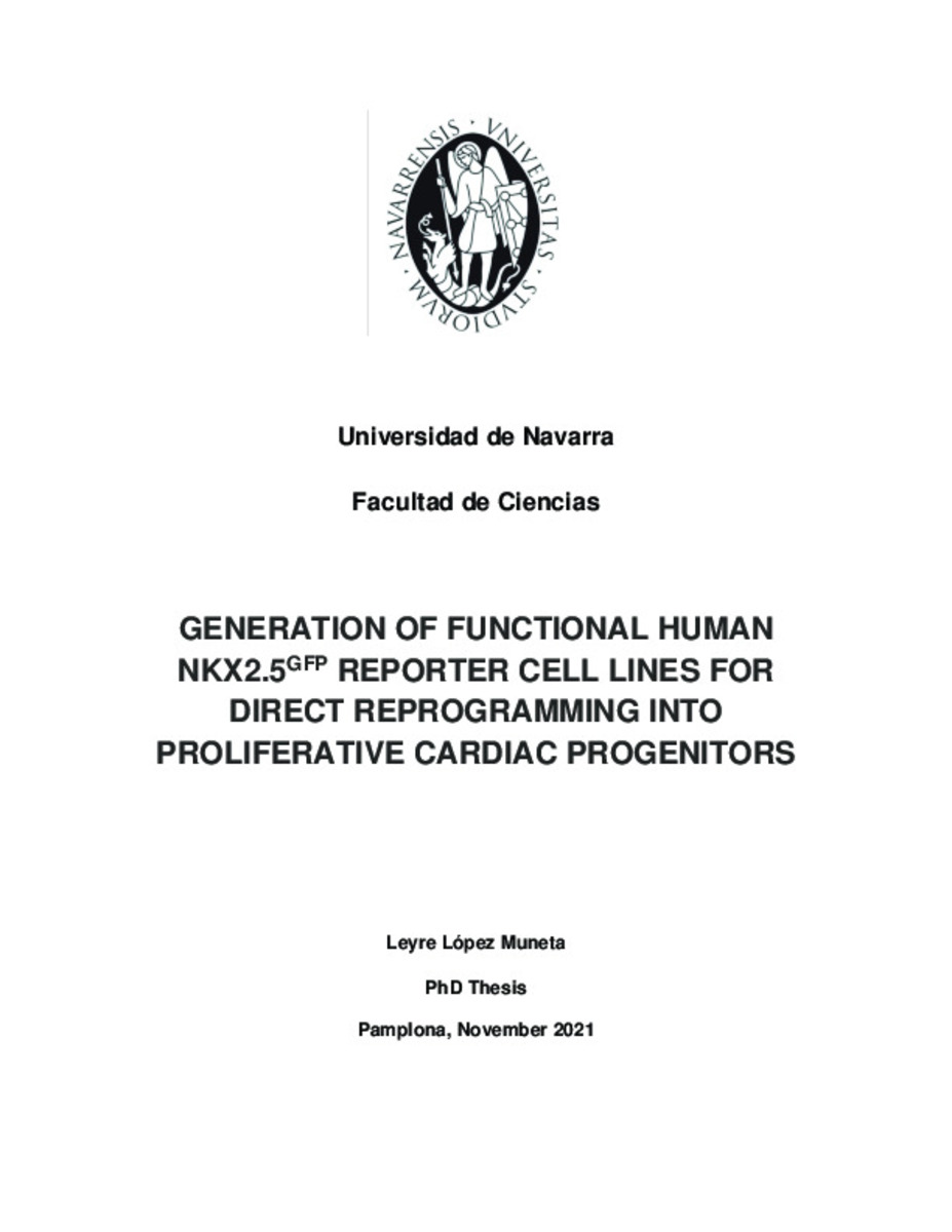
Generation of Functional Human NKX2.5GFP Reporter Cell Lines for Direct Reprogramming into Proliferative Cardiac Progenitors
Palabras clave : 
Materias Investigacion::Ciencias de la Salud::Cardiología
Cardiovascular diseases
Regeneration
Cardiac progenitors
Cellular reprogramming
Cardiac fluorescent reporter PSC lines
Fecha de publicación : 
14-dic-2021
Fecha de la defensa: 
12-nov-2021
Editorial : 
Universidad de Navarra
Cita: 
López, Leyre. "Generation of functional Human NKX2.5GFP Reporter Cell Lines for Direct Reprogramming into Proliferative Cardiac Progenitors". Carvajal, X. (dir). Tesis doctoral. Universidad de Navarra, Pamplona 2021.
Resumen
The mammalian heart is the first organ formed in the embryo and is composed of four chambers: right atrium (RA), right ventricle (RV), left atrium (LA), and left ventricle (LV), and three layers: the endocardium, the myocardium, and the pericardium. The endocardium is formed by simple squamous epithelium known as endothelium, which lines the chambers and is joined to myocardium by a thin layer of connective tissue. The pericardium is comprised of two distinct sublayers: the inner serous pericardium, which is formed by visceral pericardium or epicardium and parietal pericardium separated by pericardial cavity, and the outer fibrous pericardium made of dense connective tissue. The myocardium is the muscular layer of the heart, it is the thickest one and is located between the endocardium and the pericardium. With regard to cellular composition, the heart is principally formed by cardiomyocytes (CM), cardiac fibroblasts (cFib) , vascular smooth muscle cells (localized fundamentally within the myocardium), vascular endothelial cells (located within the myocardium and endocardium) and mesothelial cells present in the pericardium 1–3 . CM are specialized cells with a complex filament structure responsible for the control of the rhythmic beating of the heart and present heterogeneity depending on the location, morphology, and function, including atrial, ventricular, sinoatrial nodal, atrioventricular nodal, His bundle, and Purkinje fibers 4,5 .

Ficheros en este ítem:
Vista previa
Fichero
Tesis_López.pdf
Descripción
Tamaño
10.07 MB
Formato
Adobe PDF


Estadísticas e impacto
0 citas en
0 citas en

Los ítems de Dadun están protegidos por copyright, con todos los derechos reservados, a menos que se indique lo contrario.