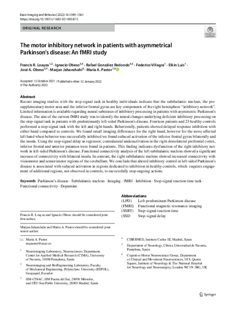
The motor inhibitory network in patients with asymmetrical Parkinson’s disease: a fMRI study
Palabras clave : 
Parkinson’s disease
Subthalamic nucleus
Imaging
fMRI
Inhibition
Stop-signal reaction time task
Functional connectivity
Dopamine
Fecha de publicación : 
2022
Editorial : 
Springer
ISSN : 
1931-7565
Nota: 
This article is licensed under a Creative Commons Attribution 4.0 International License
Cita: 
Loayza, F.R. (Francis R.); Obeso, I. (Ignacio); Gonzalez-Redondo, R. (R.); et al. "The motor inhibitory network in patients with asymmetrical Parkinson’s disease: a fMRI study". Brain Imaging and Behavior. (16), 2022, 1349 - 1361
Resumen
Recent imaging studies with the stop-signal task in healthy individuals indicate that the subthalamic nucleus, the pre-supplementary motor area and the inferior frontal gyrus are key components of the right hemisphere “inhibitory network”. Limited information is available regarding neural substrates of inhibitory processing in patients with asymmetric Parkinson’s disease. The aim of the current fMRI study was to identify the neural changes underlying deficient inhibitory processing on the stop-signal task in patients with predominantly left-sided Parkinson’s disease. Fourteen patients and 23 healthy controls performed a stop-signal task with the left and right hands. Behaviorally, patients showed delayed response inhibition with either hand compared to controls. We found small imaging differences for the right hand, however for the more affected left hand when behavior was successfully inhibited we found reduced activation of the inferior frontal gyrus bilaterally and the insula. Using the stop-signal delay as regressor, contralateral underactivation in the right dorsolateral prefrontal cortex, inferior frontal and anterior putamen were found in patients. This finding indicates dysfunction of the right inhibitory network in left-sided Parkinson’s disease. Functional connectivity analysis of the left subthalamic nucleus showed a significant increase of connectivity with bilateral insula. In contrast, the right subthalamic nucleus showed increased connectivity with visuomotor and sensorimotor regions of the cerebellum. We conclude that altered inhibitory control in left-sided Parkinson’s disease is associated with reduced activation in regions dedicated to inhibition in healthy controls, which requires engagement of additional regions, not observed in controls, to successfully stop ongoing actions.

Ficheros en este ítem:
Vista previa
Fichero
s11682-021-00587-5.pdf
Descripción
Tamaño
1.53 MB
Formato
Adobe PDF


Estadísticas e impacto
0 citas en
0 citas en

Los ítems de Dadun están protegidos por copyright, con todos los derechos reservados, a menos que se indique lo contrario.