Full metadata record
| DC Field | Value | Language |
|---|---|---|
| dc.creator | Domingo, C. (Carles) | - |
| dc.creator | Lagares, J.I. (Juan Ignacio) | - |
| dc.creator | Romero-Expósito, M. (Maite) | - |
| dc.creator | Nieto, B. (Beatriz) | - |
| dc.creator | Nieto-Camero, J.J. (Jaime J.) | - |
| dc.creator | Terrón, J.A. (José Antonio) | - |
| dc.creator | Irazola, L. (Leticia) | - |
| dc.creator | Dasu, A. (Alexandru) | - |
| dc.creator | Sánchez-Doblado, F. (Francisco) | - |
| dc.date.accessioned | 2022-08-11T09:47:58Z | - |
| dc.date.available | 2022-08-11T09:47:58Z | - |
| dc.date.issued | 2022 | - |
| dc.identifier.citation | Domingo, C.; Lagares, J. I.; Romero-Expósito, M.; et al. "Peripheral organ equivalent dose estimation procedure in proton therapy". Frontiers in Oncology. 12, 2022, 882476 | es |
| dc.identifier.issn | 2234-943X | - |
| dc.identifier.uri | https://hdl.handle.net/10171/63904 | - |
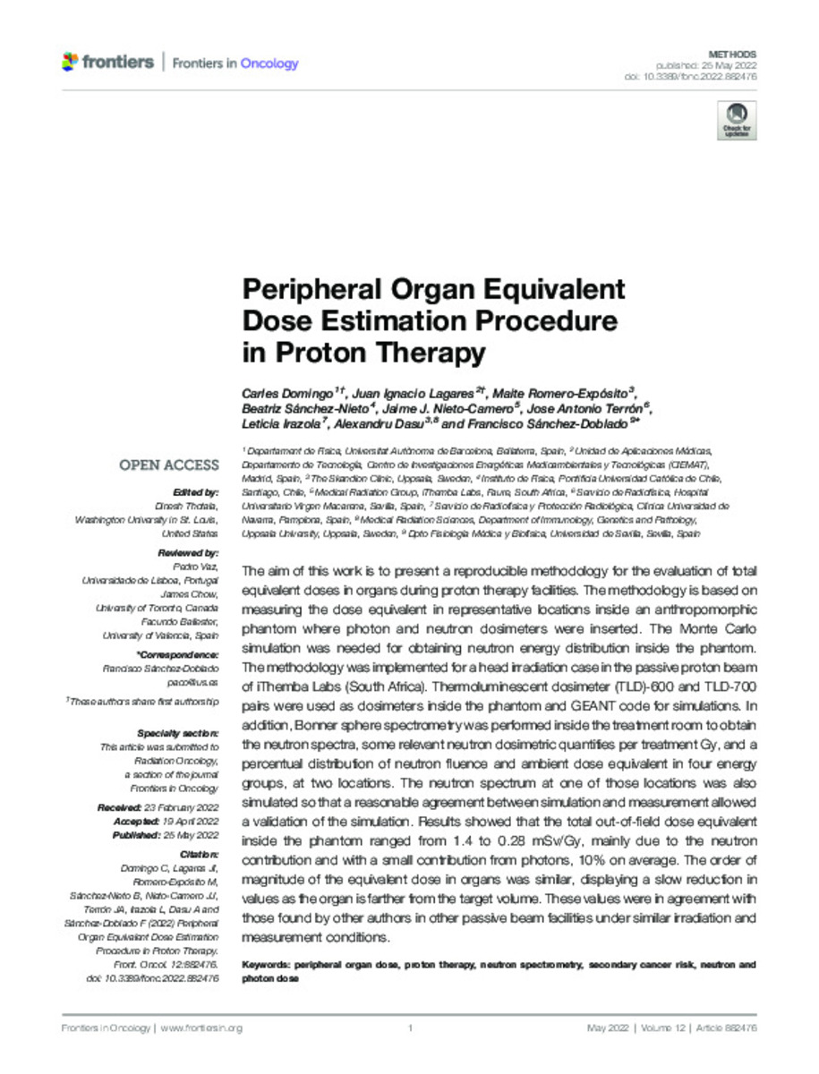
| dc.description.abstract | The aim of this work is to present a reproducible methodology for the evaluation of total equivalent doses in organs during proton therapy facilities. The methodology is based on measuring the dose equivalent in representative locations inside an anthropomorphic phantom where photon and neutron dosimeters were inserted. The Monte Carlo simulation was needed for obtaining neutron energy distribution inside the phantom. The methodology was implemented for a head irradiation case in the passive proton beam of iThemba Labs (South Africa). Thermoluminescent dosimeter (TLD)-600 and TLD-700 pairs were used as dosimeters inside the phantom and GEANT code for simulations. In addition, Bonner sphere spectrometry was performed inside the treatment room to obtain the neutron spectra, some relevant neutron dosimetric quantities per treatment Gy, and a percentual distribution of neutron fluence and ambient dose equivalent in four energy groups, at two locations. The neutron spectrum at one of those locations was also simulated so that a reasonable agreement between simulation and measurement allowed a validation of the simulation. Results showed that the total out-of-field dose equivalent inside the phantom ranged from 1.4 to 0.28 mSv/Gy, mainly due to the neutron contribution and with a small contribution from photons, 10% on average. The order of magnitude of the equivalent dose in organs was similar, displaying a slow reduction in values as the organ is farther from the target volume. These values were in agreement with those found by other authors in other passive beam facilities under similar irradiation and measurement conditions. | - |
| dc.description.sponsorship | This work has been partially carried out on the ACME cluster, which is owned by CIEMAT and funded by the Spanish Ministry of Economy and Competitiveness project CODEC2 (TIN2015- 63562-R) with FEDER funds as well as supported by the CYTED-co-founded RICAP Network (517RT0529). MR-E acknowledges funding from Euratom’s research and innovation programme 2019-20 under grant agreement no. 945196. BS-N acknowledges project Fondecyt N1181133. | - |
| dc.language.iso | en | - |
| dc.rights | info:eu-repo/semantics/openAccess | - |
| dc.subject | Peripheral organ dose | - |
| dc.subject | Proton therapy | - |
| dc.subject | Neutron spectrometry | - |
| dc.subject | Secondary cancer risk | - |
| dc.subject | Neutron and photon dose | - |
| dc.title | Peripheral organ equivalent dose estimation procedure in proton therapy | - |
| dc.type | info:eu-repo/semantics/article | - |
| dc.description.note | This is an open-access article distributed under the terms of the Creative Commons Attribution License (CC BY) | - |
| dc.identifier.doi | 10.3389/fonc.2022.882476 | - |
| dadun.citation.publicationName | Frontiers in Oncology | - |
| dadun.citation.startingPage | 882476 | - |
| dadun.citation.volume | 12 | - |
Files in This Item:
Statistics and impact
Items in Dadun are protected by copyright, with all rights reserved, unless otherwise indicated.


