Full metadata record
| DC Field | Value | Language |
|---|---|---|
| dc.creator | Scully, P.R. (Paul R.) | - |
| dc.creator | Bastarrika, G. (Gorka) | - |
| dc.creator | Moon, J.C. (James C.) | - |
| dc.creator | Treibel, T.A. (Thomas A.) | - |
| dc.date.accessioned | 2023-04-05T12:52:59Z | - |
| dc.date.available | 2023-04-05T12:52:59Z | - |
| dc.date.issued | 2018 | - |
| dc.identifier.citation | Scully, P.R. (Paul R.); Bastarrika, G. (Gorka); Moon, J.C. (James C.); et al. "Myocardial extracellular volume quantification by cardiovascularagn magnetic resonance and computed tomography". Current cardiology reports. 20 (15), 2018, | es_ES |
| dc.identifier.issn | 1523-3782 | - |
| dc.identifier.uri | https://hdl.handle.net/10171/65901 | - |
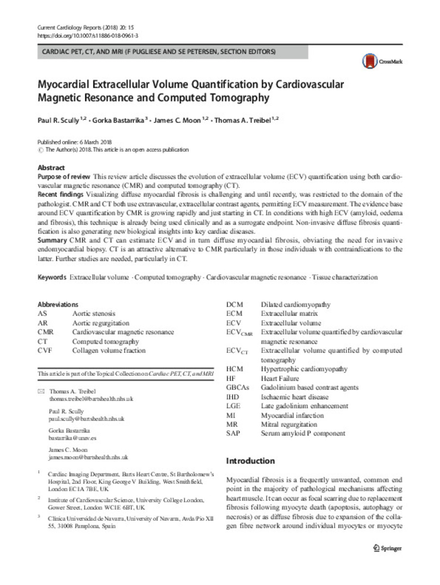
| dc.description.abstract | Purpose of review This review article discusses the evolution of extracellular volume (ECV) quantification using both cardiovascular magnetic resonance (CMR) and computed tomography (CT). Recent findings Visualizing diffuse myocardial fibrosis is challenging and until recently, was restricted to the domain of the pathologist. CMR and CT both use extravascular, extracellular contrast agents, permitting ECV measurement. The evidence base around ECV quantification by CMR is growing rapidly and just starting in CT. In conditions with high ECV (amyloid, oedema and fibrosis), this technique is already being used clinically and as a surrogate endpoint. Non-invasive diffuse fibrosis quantification is also generating new biological insights into key cardiac diseases. Summary CMR and CT can estimate ECV and in turn diffuse myocardial fibrosis, obviating the need for invasive endomyocardial biopsy. CT is an attractive alternative to CMR particularly in those individuals with contraindications to the latter. Further studies are needed, particularly in CT. | es_ES |
| dc.description.sponsorship | Paul R. Scully is supported by a British Heart Foundation Clinical Research Training Fellowship (FS/16/31/32185). James C. Moon is directly and indirectly supported by the University College London Hospitals NIHR Biomedical Research Centre and Biomedical Research Unit at Barts Hospital, respectively. Thomas Treibel was supported by doctoral research fellowship from the National Institute of Health Research (NIHR; DRF-2013-06-102). | es_ES |
| dc.language.iso | eng | es_ES |
| dc.publisher | Springer | es_ES |
| dc.rights | info:eu-repo/semantics/openAccess | es_ES |
| dc.subject | Extracellular volume | es_ES |
| dc.subject | Computed tomography | es_ES |
| dc.subject | Cardiovascular magnetic resonance | es_ES |
| dc.subject | Tissue characterization | es_ES |
| dc.title | Myocardial extracellular volume quantification by cardiovascularagn magnetic resonance and computed tomography | es_ES |
| dc.type | info:eu-repo/semantics/article | es_ES |
| dc.description.note | This article is distributed under the terms of the Creative Commons Attribution 4.0 International License (http:// creativecommons.org/licenses/by/4.0/), which permits unrestricted use, distribution, and reproduction in any medium, provided you give appropriate credit to the original author(s) and the source, provide a link to the Creative Commons license, and indicate if changes were made. | es_ES |
| dc.identifier.doi | 10.1007/s11886-018-0961-3 | - |
| dadun.citation.number | 15 | es_ES |
| dadun.citation.publicationName | Current cardiology reports | es_ES |
| dadun.citation.volume | 20 | es_ES |
| dc.identifier.pmid | 29511861 | - |
Files in This Item:
Statistics and impact
Items in Dadun are protected by copyright, with all rights reserved, unless otherwise indicated.


