Full metadata record
| DC Field | Value | Language |
|---|---|---|
| dc.creator | Stummer, W. (Walter) | - |
| dc.creator | Koch, R. (Raphael) | - |
| dc.creator | Diez-Valle, R. (Ricardo) | - |
| dc.creator | Roberts, D.W. (David W.) | - |
| dc.creator | Sanai, N. (Nadar) | - |
| dc.creator | Kalkanis, S. (Steve) | - |
| dc.creator | Hadjipanayis, C.G. (Constantinos G.) | - |
| dc.creator | Suero-Molina, E. (Eric) | - |
| dc.date.accessioned | 2021-11-04T08:41:18Z | - |
| dc.date.available | 2021-11-04T08:41:18Z | - |
| dc.date.issued | 2019 | - |
| dc.identifier.citation | Stummer, W. (Walter); Koch, R. (Raphael); Diez-Valle, R. (Ricardo); et al. "Intraoperative fluorescence diagnosis in the brain: a systematic review and suggestions for future standards on reporting diagnostic accuracy and clinical utility". Acta Neurochirurgica. 161, 2019, 2083 - 2098 | es_ES |
| dc.identifier.issn | 0001-6268 | - |
| dc.identifier.uri | https://hdl.handle.net/10171/62318 | - |
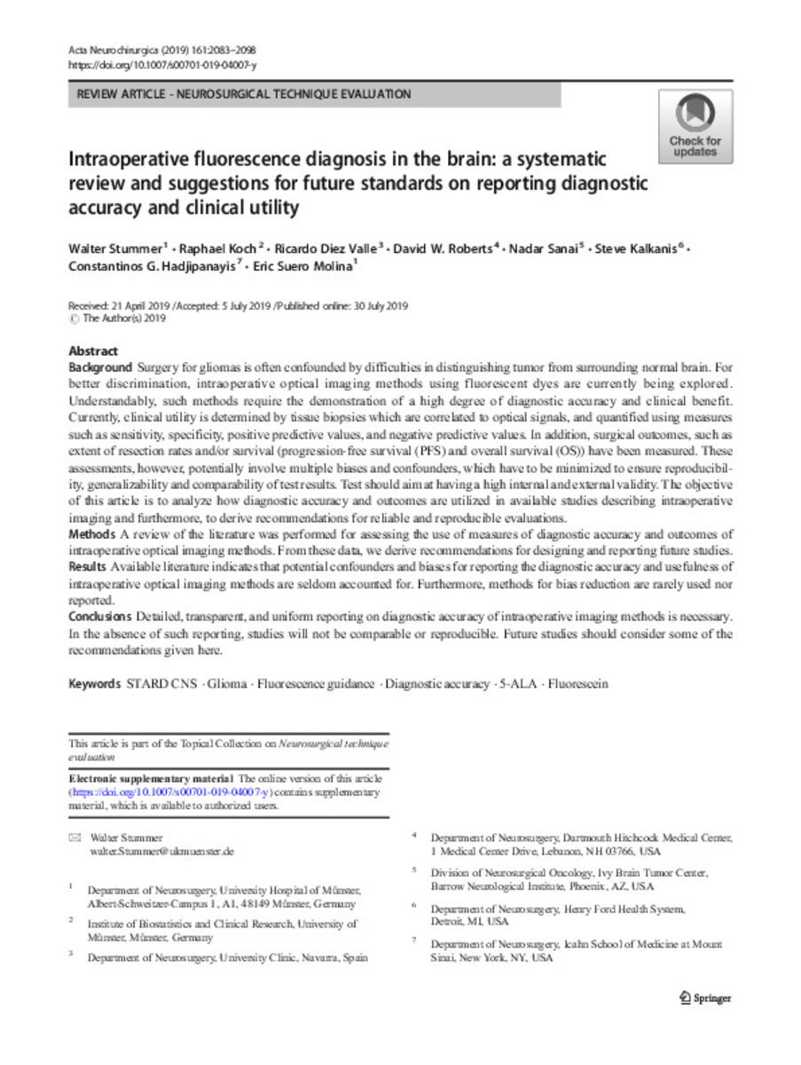
| dc.description.abstract | Background Surgery for gliomas is often confounded by difficulties in distinguishing tumor from surrounding normal brain. For better discrimination, intraoperative optical imaging methods using fluorescent dyes are currently being explored. Understandably, such methods require the demonstration of a high degree of diagnostic accuracy and clinical benefit. Currently, clinical utility is determined by tissue biopsies which are correlated to optical signals, and quantified using measures such as sensitivity, specificity, positive predictive values, and negative predictive values. In addition, surgical outcomes, such as extent of resection rates and/or survival (progression-free survival (PFS) and overall survival (OS)) have been measured. These assessments, however, potentially involve multiple biases and confounders, which have to be minimized to ensure reproducibility, generalizability and comparability of test results. Test should aim at having a high internal and external validity. The objective of this article is to analyze how diagnostic accuracy and outcomes are utilized in available studies describing intraoperative imaging and furthermore, to derive recommendations for reliable and reproducible evaluations. Methods A review of the literature was performed for assessing the use of measures of diagnostic accuracy and outcomes of intraoperative optical imaging methods. From these data, we derive recommendations for designing and reporting future studies. Results Available literature indicates that potential confounders and biases for reporting the diagnostic accuracy and usefulness of intraoperative optical imaging methods are seldom accounted for. Furthermore, methods for bias reduction are rarely used nor reported. Conclusions Detailed, transparent, and uniform reporting on diagnostic accuracy of intraoperative imaging methods is necessary. In the absence of such reporting, studies will not be comparable or reproducible. Future studies should consider some of the recommendations given here. | es_ES |
| dc.language.iso | eng | es_ES |
| dc.publisher | Springer Science and Business Media LLC | es_ES |
| dc.rights | info:eu-repo/semantics/openAccess | es_ES |
| dc.subject | STARD CNS | es_ES |
| dc.subject | Glioma | es_ES |
| dc.subject | Fluorescence guidance | es_ES |
| dc.subject | Diagnostic accuracy | es_ES |
| dc.subject | 5-ALA | es_ES |
| dc.subject | Fluorescein | es_ES |
| dc.title | Intraoperative fluorescence diagnosis in the brain: a systematic review and suggestions for future standards on reporting diagnostic accuracy and clinical utility | es_ES |
| dc.type | info:eu-repo/semantics/article | es_ES |
| dc.description.note | Open Access This article is distributed under the terms of the Creative Commons Attribution 4.0 International License | es_ES |
| dc.identifier.doi | 10.1007/s00701-019-04007-y | - |
| dadun.citation.endingPage | 2098 | es_ES |
| dadun.citation.publicationName | Acta Neurochirurgica | es_ES |
| dadun.citation.startingPage | 2083 | es_ES |
| dadun.citation.volume | 161 | es_ES |
Files in This Item:
Statistics and impact
Items in Dadun are protected by copyright, with all rights reserved, unless otherwise indicated.


