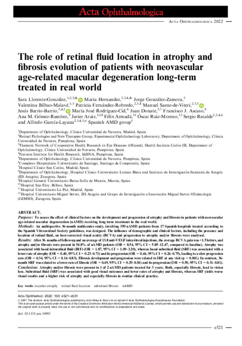
The role of retinal fluid location in atrophy and fibrosis evolution of patients with neovascular age-related macular degeneration long-term treated in real world
Palabras clave : 
Macular atrophy
Retinal fluid location
Subretinal fibrosis
nAMD
Fecha de publicación : 
2022
Editorial : 
Wiley
ISSN : 
1755-375X
Nota: 
This is an open access article under the terms of the Creative Commons Attribution-NonCommercial-NoDerivs License
Cita: 
Llorente-González, S. (Sara); Hernandez, M. (María); González-Zamora, J. (Jorge); et al. "The role of retinal fluid location in atrophy and fibrosis evolution of patients with neovascular age-related macular degeneration long-term treated in real world". Acta Ophthalmologica. 100 (2), 2022, 521 - 531
Resumen
Purpose: To assess the effect of clinical factors on the development and progression of atrophy and fibrosis in patients with neovascular age-related macular degeneration (nAMD) receiving long-term treatment in the real world. Methods: An ambispective 36-month multicentre study, involving 359 nAMD patients from 17 Spanish hospitals treated according to the Spanish Vitreoretinal Society guidelines, was designed. The influence of demographic and clinical factors, including the presence and location of retinal fluid, on best-corrected visual acuity (BCVA) and progression to atrophy and/or fibrosis were analysed. Results: After 36 months of follow-up and an average of 13.8 anti-VEGF intravitreal injections, the average BCVA gain was +1.5 letters, and atrophy and/or fibrosis were present in 54.8% of nAMD patients (OR = 8.54, 95% CI = 5.85-12.47, compared to baseline). Atrophy was associated with basal intraretinal fluid (IRF) (OR = 1.87, 95% CI = 1.09-3.20), whereas basal subretinal fluid (SRF) was associated with a lower rate of atrophy (OR = 0.40, 95% CI = 0.23-0.71) and its progression (OR = 0.44, 95% CI = 0.26-0.75), leading to a slow progression rate (OR = 0.34, 95% CI = 0.14-0.83). Fibrosis development and progression were related to IRF at any visit (p < 0.001). In contrast, 36-month SRF was related to a lower rate of fibrosis (OR = 0.49, 95% CI = 0.29-0.81) and its progression (OR = 0.50, 95% CI = 0.31-0.81). Conclusion: Atrophy and/or fibrosis were present in 1 of 2 nAMD patients treated for 3 years. Both, especially fibrosis, lead to vision loss. Subretinal fluid (SRF) was associated with good visual outcomes and lower rates of atrophy and fibrosis, whereas IRF yields worse visual results and a higher risk of atrophy and especially fibrosis in routine clinical practice.

Ficheros en este ítem:
Vista previa
Fichero
Acta Ophthalmologica - 2021 - Llorente‐Gonz lez - The role of retinal fluid location in atrophy and fibrosis evolution of.pdf
Descripción
Tamaño
1.13 MB
Formato
Adobe PDF


Estadísticas e impacto
0 citas en
0 citas en

Los ítems de Dadun están protegidos por copyright, con todos los derechos reservados, a menos que se indique lo contrario.