Full metadata record
| DC Field | Value | Language |
|---|---|---|
| dc.creator | Rodríguez-Marco, N.A. (Nelson Arturo) | - |
| dc.creator | Caicedo-Zamudio, C. (Carlos) | - |
| dc.creator | Solanas-Álava, S. (S.) | - |
| dc.creator | Gil-Arnaiz, I. (Irene) | - |
| dc.creator | Córdoba-Iturriagagoitia, A. (Alicia) | - |
| dc.creator | Andonegui-Navarro, J. (J.) | - |
| dc.date.accessioned | 2023-02-17T12:36:18Z | - |
| dc.date.available | 2023-02-17T12:36:18Z | - |
| dc.date.issued | 2014 | - |
| dc.identifier.citation | Rodríguez-Marco, N.A. (Nelson Arturo); Caicedo-Zamudio, C. (Carlos); Solanas-Álava, S. (S.); et al. "PET/TC de cuerpo completo para la detección de metástasis de melanoma coroideo". Anales del Sistema Sanitario de Navarra. 37 (2), 2014, 293 - 298 | es |
| dc.identifier.issn | 1137-6840 | - |
| dc.identifier.uri | https://hdl.handle.net/10171/65500 | - |
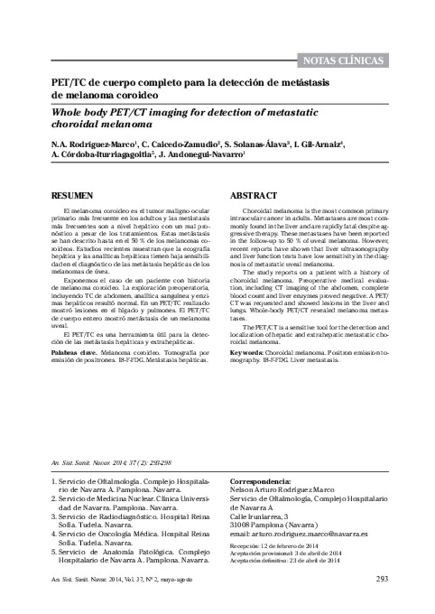
| dc.description.abstract | El melanoma coroideo es el tumor maligno ocular primario más frecuente en los adultos y las metástasis más frecuentes son a nivel hepático con un mal pronóstico a pesar de los tratamientos. Estas metástasis se han descrito hasta en el 50 % de los melanomas coroideos. Estudios recientes muestran que la ecografía hepática y las analíticas hepáticas tienen baja sensibilidad en el diagnóstico de las metástasis hepáticas de los melanomas de úvea. Exponemos el caso de un paciente con historia de melanoma coroideo. La exploración preoperatoria, incluyendo TC de abdomen, analítica sanguínea y enzimas hepáticos resultó normal. En un PET/TC realizado mostró lesiones en el hígado y pulmones. El PET/TC de cuerpo entero mostró metástasis de un melanoma uveal. El PET/TC es una herramienta útil para la detección de las metástasis hepáticas y extrahepáticas. | es_ES |
| dc.description.abstract | Choroidal melanoma is the most common primary intraocular cancer in adults. Metastases are most commonly found in the liver and are rapidly fatal despite aggressive therapy. These metastases have been reported in the follow-up to 50 % of uveal melanoma. However, recent reports have shown that liver ultrasonography and liver function tests have low sensitivity in the diagnosis of metastatic uveal melanoma. The study reports on a patient with a history of choroidal melanoma. Preoperative medical evaluation, including CT imaging of the abdomen, complete blood count and liver enzymes proved negative. A PET/ CT was requested and showed lesions in the liver and lungs. Whole-body PET/CT revealed melanoma metastases. The PET/CT is a sensitive tool for the detection and localization of hepatic and extrahepatic metastatic choroidal melanoma. | es_ES |
| dc.language.iso | spa | es_ES |
| dc.rights | info:eu-repo/semantics/openAccess | es_ES |
| dc.subject | Melanoma coroideo | es_ES |
| dc.subject | Tomografía por emisión de positrones | es_ES |
| dc.subject | 18-F-FDG | es_ES |
| dc.subject | Metástasis hepáticas | es_ES |
| dc.subject | Choroidal melanoma | es_ES |
| dc.subject | Positron emission tomography | es_ES |
| dc.subject | Liver metastasis | es_ES |
| dc.title | PET/TC de cuerpo completo para la detección de metástasis de melanoma coroideo | es_ES |
| dc.title.alternative | Whole body PET/CT imaging for detection of metastatic choroidal melanoma | es_ES |
| dc.type | info:eu-repo/semantics/article | es_ES |
| dc.publisher.place | Navarra | es_ES |
| dc.description.note | CC BY NC | es_ES |
| dc.identifier.doi | 10.4321/S1137-66272014000200013 | - |
| dadun.citation.endingPage | 298 | es_ES |
| dadun.citation.number | 2 | es_ES |
| dadun.citation.publicationName | Anales del Sistema Sanitario de Navarra | es_ES |
| dadun.citation.startingPage | 293 | es_ES |
| dadun.citation.volume | 37 | es_ES |
| dc.identifier.pmid | 25189988 | - |
Files in This Item:
Statistics and impact
Items in Dadun are protected by copyright, with all rights reserved, unless otherwise indicated.


